
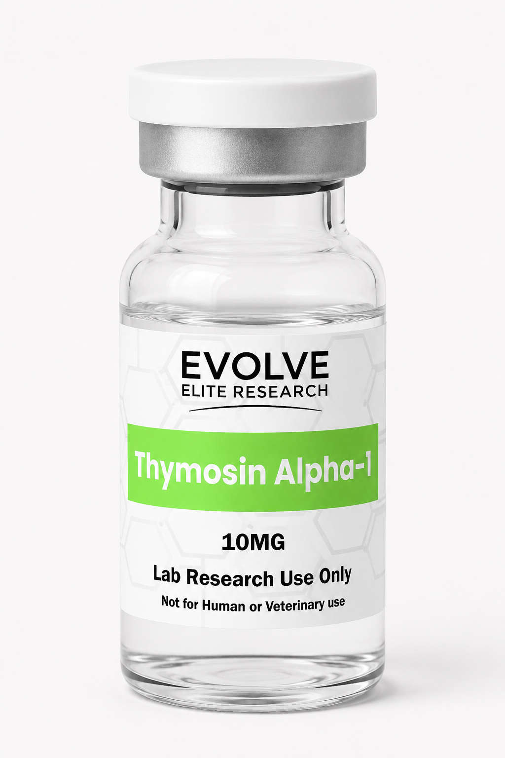
Thymosin Alpha-1 10MG
$67.00
Technical Specifications
- Scientific Research Name: Thymosin Alpha-1
- Alternate Name: Thymalfasin (TA1)
- Research Description: 28-amino-acid synthetic thymic peptide studied in immune-signaling models
- Compound Classification: Synthetic peptide
- Primary Research Areas: Dendritic-cell biology, T-cell signaling, pattern-recognition pathways and cytokine research
- Research Format: 10MG lyophilized research material
- Intended Use: Laboratory research only
For Laboratory Research Use Only.Not intended for human or veterinary administration.
In stock
🔬 For Laboratory Research Use Only.
Not for human consumption, medical, veterinary, or household use.
By purchasing you agree to our
Terms and
Refund Policy.
Ideal for replicate studies, concentration-response assays and extended research workflows
Synthetic analogue of the naturally occurring thymic peptide Thymosin Alpha-1
Studied in dendritic cell, T-cell and innate immune communication models
Shipped from our Texas facility with fast U.S. order processing
Lyophilized peptide supplied for controlled laboratory research involving immune-signaling pathways, peptide–cell interactions, receptor-associated activity, and experimental immunology models.
Thymosin Alpha-1 10MG Research Overview
Thymosin Alpha-1 10MG is a laboratory research material consisting of a 28-amino-acid peptide corresponding to a naturally occurring fragment derived from prothymosin alpha. Also commonly abbreviated as TA1 or Tα1 in scientific literature, the peptide has been investigated in laboratory models involving innate and adaptive immune signaling, dendritic-cell activity, T-cell differentiation, cytokine regulation, pathogen-response pathways, and immune-system homeostasis.
The Thymosin Alpha-1 sequence contains several acidic amino-acid residues and has a reported molecular weight of approximately 3108.30 g/mol. Its structure and biochemical properties have made it a subject of experimental research examining how short endogenous peptides may influence cellular communication within immune-associated systems.
In controlled research environments, Thymosin Alpha-1 may be evaluated through cell-culture systems, receptor-signaling assays, gene-expression studies, cytokine panels, immune-cell differentiation models, biochemical characterization, and comparative peptide research. Experimental design should account for peptide concentration, assay format, biological matrix, exposure interval, control conditions, analytical sensitivity, and the stability requirements of the selected model.
This 10MG format provides researchers with a larger quantity of material for projects requiring repeat assays, replicate testing, multi-condition experiments, analytical verification, or extended laboratory workflows. The product is supplied as a lyophilized powder for research use only.
Technical Specifications
Thymosin Alpha-1 10MG
TA1, Tα1, Thymosin α1
Synthetic research peptide
10MG per vial
Lyophilized powder
28 amino acids
62304-98-7
16132342
3108.30 g/mol
C129H215N33O55
Immune-signaling peptide research
Laboratory research only
Molecular and Sequence Data
Thymosin Alpha-1 is composed of 28 amino-acid residues. Its relatively short sequence allows researchers to evaluate the peptide through analytical methods commonly used for synthetic peptide characterization, including chromatographic separation, mass analysis, sequence verification, and stability testing.
Ser-Asp-Ala-Ala-Val-Asp-Thr-Ser-Ser-Glu-Ile-Thr-Thr-Lys-Asp-Leu-Lys-Glu-Lys-Lys-Glu-Val-Val-Glu-Glu-Ala-Glu-Asn
The sequence contains multiple glutamic-acid and aspartic-acid residues, contributing to the peptide’s acidic character. Lysine residues introduce positively charged side chains, while serine and threonine residues provide polar hydroxyl groups that may influence peptide conformation, solvent interaction, and experimental behavior.
Sequence composition can affect chromatographic retention, ionization behavior, solubility, aggregation potential, and susceptibility to degradation. Researchers should therefore interpret analytical and biological results in relation to the full sequence rather than relying solely on nominal peptide concentration.
Aspartic-acid and glutamic-acid residues contribute negatively charged side chains under many common experimental conditions.
Lysine residues provide positively charged functional groups that may influence intermolecular interactions and ionization patterns.
Serine, threonine, asparagine, and other polar residues affect hydration, hydrogen bonding, and peptide–solvent interactions.
Alanine, valine, leucine, and isoleucine contribute hydrophobic character that may influence local structural organization.
Scientific Background
Thymosin Alpha-1 was originally identified through research involving thymic peptide fractions. It was later characterized as a specific peptide sequence associated with prothymosin alpha, a larger intracellular protein. Research involving Thymosin Alpha-1 expanded as investigators examined the influence of thymic peptides on immune-cell development, immune communication, and host-response signaling.
The thymus is closely associated with T-lymphocyte development and selection. This relationship led researchers to investigate thymic peptides as possible molecular participants in immune-cell maturation and signaling. Thymosin Alpha-1 became one of the most extensively studied peptides within this category because its sequence could be synthesized, characterized, and evaluated in reproducible laboratory systems.
Contemporary research does not treat Thymosin Alpha-1 as a simple one-receptor ligand with a single downstream outcome. Instead, the peptide is examined within complex biological networks involving pattern-recognition signaling, antigen presentation, transcriptional regulation, cytokine communication, immune-cell differentiation, and interactions between innate and adaptive responses.
Reported experimental effects may vary substantially according to the cell type, biological species, activation state, model design, peptide sample condition, concentration, exposure duration, and assay endpoint. Results from one experimental system should not automatically be generalized to another.
Prothymosin Alpha and Peptide Origin
Prothymosin alpha is a larger acidic protein involved in multiple intracellular processes. Thymosin Alpha-1 corresponds to a defined N-terminal sequence associated with prothymosin alpha. Understanding this relationship is important because the isolated 28-amino-acid peptide and the full-length precursor protein are not interchangeable research materials.
The isolated peptide may be studied as an individual molecular entity under controlled conditions, while the precursor protein participates in broader cellular systems. Researchers should clearly identify which material is being evaluated and avoid attributing findings from full-length prothymosin-alpha research directly to synthetic Thymosin Alpha-1 without experimental support.
Synthetic production allows the peptide sequence to be investigated independently of complex biological extracts. This supports controlled comparison of concentration, purity, structural identity, analytical response, and biological activity across experimental conditions.
Innate and Adaptive Immune Research
The immune system is frequently divided into innate and adaptive components for experimental analysis. Innate immunity includes rapid-response mechanisms involving pattern-recognition receptors, macrophages, monocytes, dendritic cells, natural killer cells, and soluble signaling molecules. Adaptive immunity includes antigen-specific T-cell and B-cell responses, immunological memory, and antibody-associated processes.
Thymosin Alpha-1 is of interest because research has explored its potential interaction with both branches. In experimental systems, the peptide has been evaluated in relation to dendritic-cell maturation, antigen presentation, T-cell signaling, natural killer-cell activity, cytokine production, and communication between innate sensing and adaptive immune responses.
This does not mean that every model produces the same response. Immune activity is highly dependent on baseline activation state, cell composition, receptor expression, inflammatory environment, antigen exposure, and the timing of measurement. Appropriate negative controls, stimulated controls, vehicle controls, and reference compounds are therefore essential.
Experimental systems may examine macrophages, dendritic cells, monocytes, natural killer cells, pattern-recognition signaling, or early cytokine responses.
Research may evaluate T-cell differentiation, activation markers, antigen-specific signaling, regulatory pathways, or lymphocyte communication.
Researchers may investigate how innate sensing influences antigen presentation and downstream adaptive immune activity.
Some experimental designs focus on immune balance, signaling normalization, or differences between resting and activated cellular states.
Pattern-Recognition Receptor Research
Pattern-recognition receptors detect conserved molecular features associated with microbes, cellular stress, or tissue disruption. Toll-like receptors are among the most frequently studied pattern-recognition receptor families. They participate in signaling pathways that can activate transcription factors, cytokine production, antigen-presentation programs, and other components of innate immune defense.
Thymosin Alpha-1 has been investigated in experimental models involving Toll-like receptor-associated pathways. Some studies have examined relationships with TLR2, TLR9, MyD88-dependent signaling, dendritic-cell function, and downstream transcriptional responses.
These interactions should be described carefully. Thymosin Alpha-1 is not universally established as a conventional high-affinity ligand for a single Toll-like receptor in the same manner as a defined microbial agonist. Research more commonly evaluates how the peptide may influence receptor-associated signaling networks, cellular responsiveness, or immune communication.
Experimental assessment may include receptor-expression analysis, reporter assays, phosphorylation measurements, transcription-factor localization, cytokine release, gene-expression profiling, and comparison with established receptor agonists or antagonists.
Dendritic-Cell Research
Dendritic cells act as an important bridge between innate sensing and adaptive immune activation. These cells detect environmental signals, process antigens, migrate between tissues, and present peptide fragments to T lymphocytes through major histocompatibility complexes.
Thymosin Alpha-1 has been evaluated in dendritic-cell research involving cellular maturation, antigen presentation, cytokine production, pattern-recognition signaling, and interaction with T-cell populations. Researchers may compare immature and mature dendritic-cell phenotypes after controlled exposure to the peptide.
Common experimental markers may include surface proteins associated with antigen presentation, co-stimulation, maturation, and migration. Flow cytometry, microscopy, gene-expression analysis, cytokine assays, and functional co-culture systems may be used to assess changes.
Dendritic-cell responses can differ according to tissue source, differentiation protocol, species, maturation stimulus, culture media, and baseline activation state. A well-controlled study should document these factors and avoid interpreting a single marker as proof of a complete functional phenotype.
T-Cell Signaling and Differentiation Research
T lymphocytes are central to adaptive immune research. Their functions include recognition of antigen-associated signals, production of signaling molecules, regulation of other immune populations, direct cytotoxic activity, and formation of memory-cell populations.
Thymosin Alpha-1 has been studied in relation to T-cell maturation, differentiation, activation, proliferation, and cytokine-associated behavior. Experimental models may evaluate helper T-cell populations, cytotoxic T cells, regulatory T cells, memory-associated phenotypes, or the balance between different differentiation programs.
T-cell responses depend on multiple signals. These may include antigen-receptor engagement, co-stimulatory molecules, cytokine exposure, metabolic state, and inhibitory receptor activity. Thymosin Alpha-1 should therefore be evaluated as one component of a larger experimental system rather than as an isolated determinant of T-cell behavior.
Natural Killer Cell Research
Natural killer cells are innate lymphoid cells studied for their ability to recognize and respond to stressed, infected, or transformed cells without requiring the same antigen-specific priming used by conventional T cells.
Laboratory research involving Thymosin Alpha-1 has explored natural killer-cell activity, signaling, cytokine communication, and interactions with other immune-cell populations. Experimental models may assess changes in surface-marker expression, cytotoxicity-associated proteins, target-cell interaction, degranulation, or cytokine production.
Natural killer-cell assays should include careful control of effector-to-target ratios, target-cell identity, incubation duration, viability, and baseline activity. Results may differ considerably between purified NK-cell systems and mixed peripheral immune-cell preparations.
Macrophage and Monocyte Research
Macrophages and monocytes participate in pathogen recognition, phagocytosis, cytokine production, antigen processing, tissue signaling, and inflammatory regulation. Their phenotype is highly responsive to environmental conditions.
Thymosin Alpha-1 may be evaluated in macrophage or monocyte systems to examine receptor-associated signaling, transcriptional responses, inflammatory mediators, phagocytic behavior, oxidative responses, or communication with lymphocyte populations.
Simplified labels such as pro-inflammatory and anti-inflammatory can be useful for basic discussion but do not fully capture macrophage diversity. Modern experimental analysis often relies on multi-marker panels, transcriptomics, metabolic measurements, and functional testing rather than binary classification.
Researchers should distinguish between direct peptide-associated effects and responses caused by experimental contaminants, vehicle conditions, endotoxin exposure, or changes in cell viability. Endotoxin control is particularly important in immune-cell research because small amounts can strongly influence pattern-recognition pathways.
Cytokine-Signaling Research
Cytokines are soluble signaling proteins that allow immune cells and other cell types to coordinate activity. Their functions depend on concentration, timing, receptor availability, tissue context, and interactions with other signaling molecules.
Thymosin Alpha-1 research has examined cytokine-associated responses in a variety of experimental models. Depending on the system, investigators may measure interferons, interleukins, chemokines, colony-stimulating factors, or inflammatory mediators.
Cytokine findings should not be interpreted as universally stimulatory or inhibitory. A peptide may be associated with different patterns in resting cells, pathogen-stimulated cells, tumor-associated models, or immune-suppressed systems. The timing of sample collection may also determine whether an early transient response or a later regulatory phase is observed.
ELISA-based testing may be used when a study focuses on one predefined cytokine or signaling molecule.
Bead-based or array-based platforms can measure multiple cytokines and chemokines from the same experimental sample.
Quantitative PCR or sequencing may evaluate transcriptional changes associated with cytokine production and receptor signaling.
Multiple collection intervals may help distinguish rapid signaling responses from delayed regulatory changes.
Transcription-Factor and Intracellular Signaling Research
Immune-cell activation depends on intracellular signaling networks that connect receptors at the cell surface or within intracellular compartments to changes in transcription, metabolism, protein expression, and cellular behavior.
Thymosin Alpha-1 research may involve pathways associated with nuclear factor kappa B, interferon-regulatory factors, mitogen-activated protein kinases, STAT proteins, MyD88-dependent signaling, and other immune-response networks. The specific pathway examined should be selected according to the biological model and experimental hypothesis.
Common methods include Western blotting, phospho-protein analysis, reporter-gene assays, immunofluorescence, nuclear-translocation studies, quantitative PCR, RNA sequencing, and pathway-inhibition experiments.
Correlation between peptide exposure and pathway activation does not automatically prove direct molecular binding. Receptor-blocking studies, inhibitors, genetic knockdown, knockout models, and complementary biochemical approaches can help determine whether an observed response is direct, indirect, or dependent on another signaling component.
Major Histocompatibility Complex and Antigen-Presentation Research
Antigen presentation allows immune cells to display peptide fragments through major histocompatibility complex molecules. T lymphocytes inspect these complexes through antigen receptors and may become activated when the required signaling conditions are present.
Research involving Thymosin Alpha-1 has explored antigen-presenting cells, maturation markers, co-stimulatory signaling, and communication between dendritic cells and T lymphocytes. These studies may help investigators examine whether peptide exposure changes the ability of an antigen-presenting cell to support downstream immune responses.
Experimental endpoints may include MHC expression, co-stimulatory molecules, antigen uptake, antigen processing, T-cell proliferation, cytokine production, and changes in cell–cell interaction.
Because antigen-presentation systems involve multiple cell types and signaling stages, researchers should include both antigen-dependent and antigen-independent controls. This helps separate general cellular activation from antigen-specific effects.
Interferon-Associated Research
Interferons are signaling proteins involved in cellular responses to viral material, immune activation, antigen presentation, and communication between innate and adaptive systems. Type I, type II, and type III interferons activate related but distinct receptor pathways and transcriptional programs.
Thymosin Alpha-1 has been examined in experimental systems involving interferon-associated pathways, including models where researchers evaluate whether combined signals alter gene expression, immune-cell activity, or pathogen-response markers.
Combination research requires careful interpretation because observed effects may be additive, synergistic, antagonistic, or independent. A factorial design that evaluates each material separately and in combination is preferable to comparing only an untreated group with a combined-exposure group.
Researchers may evaluate interferon-stimulated genes, STAT phosphorylation, cytokine release, antigen-presentation markers, viral-replication endpoints, or cell viability. Findings from these models remain specific to the experimental conditions used.
Pathogen-Response Research
Thymosin Alpha-1 has been included in laboratory research involving cellular responses to viral, bacterial, and fungal challenges. These studies generally focus on immune signaling, host-response pathways, pathogen-recognition mechanisms, cellular defense programs, or interactions between immune populations.
Pathogen-response research should distinguish between direct antimicrobial or antiviral activity and indirect effects mediated through host cells. A change in pathogen-associated measurements may result from altered immune signaling, cell viability, receptor expression, or experimental conditions rather than direct interaction between the peptide and the pathogen.
Useful controls may include pathogen-only conditions, peptide-only conditions, infected untreated cells, vehicle controls, known pathway activators, known inhibitors, and direct cell-free assays where appropriate.
Inflammatory-Response Research
Inflammation is a coordinated biological process involving immune-cell recruitment, vascular signaling, cytokines, chemokines, lipid mediators, transcriptional regulation, and tissue-specific responses. It can be protective, damaging, acute, chronic, localized, or systemic depending on the model.
Thymosin Alpha-1 may be studied in inflammatory models to examine whether peptide exposure influences cytokine patterns, immune-cell activation, transcription-factor signaling, oxidative responses, or communication between innate and adaptive immune systems.
Researchers should avoid reducing inflammatory activity to a single marker. A decrease in one cytokine does not necessarily indicate broad suppression, and an increase in another does not automatically demonstrate harmful activation. Multi-endpoint analysis provides a more complete interpretation.
Cell viability and metabolic status should be measured alongside inflammatory endpoints. Apparent decreases in cytokine production may result from reduced cell number or compromised viability rather than pathway-specific regulation.
Immune Homeostasis Research
Immune homeostasis refers to the regulation required to maintain protective immune capacity without uncontrolled or prolonged activation. This balance involves activating receptors, inhibitory receptors, regulatory cells, cytokine networks, tissue-derived signals, and programmed resolution pathways.
Thymosin Alpha-1 research sometimes focuses on immunomodulation rather than simple immune stimulation. In this context, investigators may examine whether peptide-associated responses differ between resting, activated, suppressed, or dysregulated experimental states.
A material described as immunomodulatory may produce context-dependent findings. The same experimental compound could be associated with increased activity in one model and reduced inflammatory signaling in another because the baseline biological conditions differ.
Researchers should therefore define the starting state of the model and avoid assuming that all immune-associated effects move in one direction.
Cell-Survival and Apoptosis Research
Immune-system research frequently includes measurement of cell survival, apoptosis, proliferation, and stress responses. These endpoints help determine whether observed changes in signaling are related to pathway regulation, altered cell number, or cytotoxicity.
Thymosin Alpha-1 may be evaluated through assays measuring membrane integrity, mitochondrial activity, caspase activation, DNA fragmentation, phosphatidylserine exposure, proliferation, or cell-cycle distribution.
Researchers should use more than one viability or apoptosis method where possible. Metabolic assays, for example, measure enzyme activity rather than cell number directly and can be influenced by changes in cellular metabolism.
Concentration-response testing is particularly useful because it helps distinguish a narrow signaling effect from nonspecific changes that appear only at high experimental concentrations.
Cancer-Immunology Research
Cancer immunology examines interactions among transformed cells, immune populations, tumor-associated signaling, antigen presentation, immune checkpoints, inflammatory mediators, and the surrounding cellular environment.
Thymosin Alpha-1 has been investigated in controlled experimental research involving immune responses within cancer-immunology models. Experimental questions may include dendritic-cell behavior, T-cell activity, natural killer cells, cytokine signaling, antigen presentation, or interactions with other research compounds.
The peptide should not be characterized as directly cytotoxic to transformed cells without supporting assay data or as producing a uniform response across cancer-immunology models. Experimental findings vary according to model type, immune composition, genetic background, culture conditions, and study design.
Researchers may use co-culture systems, organoids, immune-cell profiling, cytokine panels, target-cell assays, gene-expression analysis, or other controlled model systems. Results remain specific to the experimental conditions and endpoints used.
Immune-Checkpoint Research
Immune checkpoints are receptor–ligand systems that regulate immune activation and help limit inappropriate tissue damage. In tumor-associated models, checkpoint signaling can also influence the ability of immune cells to respond to transformed cells.
Thymosin Alpha-1 may be included in experimental combination studies involving immune-checkpoint pathways. Such research may evaluate T-cell activation, exhaustion-associated markers, cytokine release, proliferation, cytotoxicity, or changes within the immune-cell composition of a model.
Combination experiments should include each component as an individual condition. Without these controls, researchers cannot determine whether an observed response is attributable to the peptide, the checkpoint-associated material, or an interaction between them.
Findings from checkpoint research are model-dependent and should not be generalized beyond the experimental system in which they were generated.
Antigen-Response Research
Antigen-response laboratory research may examine antigen presentation, dendritic-cell maturation, T-cell responses, B-cell signaling, antibody-associated measurements, cytokine patterns, and formation of memory-associated immune populations.
Thymosin Alpha-1 has been investigated in experimental antigen-response models. Researchers may evaluate whether peptide exposure is associated with changes in the magnitude, timing, or cellular composition of an antigen-specific response.
These studies require careful differentiation among changes in antigen presentation, nonspecific immune activation, altered signaling, and differences in immune-cell survival or proliferation.
Experimental results from antigen-response models remain specific to the selected system, controls, and measured endpoints.
Fibrosis and Tissue-Signaling Research
Fibrosis research examines excessive extracellular-matrix deposition, fibroblast activity, inflammatory communication, tissue remodeling, growth-factor signaling, and interactions between resident cells and infiltrating immune populations.
Because immune signaling contributes to many fibrosis-associated models, Thymosin Alpha-1 may be studied in relation to cytokine patterns, macrophage behavior, inflammatory-cell recruitment, or immune–stromal interactions.
Researchers should distinguish between direct effects on fibroblasts and indirect effects mediated by immune-cell signaling. Co-culture systems, conditioned-media experiments, pathway inhibitors, and cell-specific assays may help separate these mechanisms.
Common endpoints may include collagen-associated measurements, extracellular-matrix genes, fibroblast activation markers, cytokines, tissue histology, and pathway-specific signaling proteins.
Oxidative-Stress Research
Oxidative stress occurs when reactive molecular species exceed a biological system’s ability to regulate or neutralize them. Immune activation, mitochondrial activity, pathogen responses, and tissue injury can all contribute to oxidative signaling.
Thymosin Alpha-1 may be evaluated in oxidative-stress models as part of broader research involving immune-cell activity, inflammatory signaling, mitochondrial function, or cellular defense pathways.
Common endpoints include reactive-oxygen-species probes, antioxidant enzymes, lipid oxidation, protein oxidation, glutathione status, mitochondrial membrane potential, and cell survival.
Fluorescent oxidative-stress probes can be affected by cell number, probe loading, light exposure, and nonspecific chemical reactions. Appropriate controls and complementary assays are recommended.
Potential Research Applications
Evaluation of intracellular pathways associated with immune-cell activation, regulation, and communication.
Investigation of maturation, antigen presentation, co-stimulatory markers, and interaction with lymphocyte populations.
Study of differentiation, activation markers, proliferation, cytokine production, and regulatory signaling.
Analysis of innate lymphoid-cell signaling, target-cell interaction, degranulation, and cytokine-associated activity.
Evaluation of Toll-like receptor-associated signaling and downstream transcriptional responses.
Measurement of individual or multiplexed immune-signaling molecules across controlled experimental conditions.
Study of host-cell signaling after exposure to viral, bacterial, fungal, or pathogen-associated stimuli.
Investigation of immune-cell communication, antigen presentation, target-cell responses, and combination research.
Analysis of cytokines, chemokines, transcription factors, cell viability, and immune regulatory processes.
Comparative testing alongside antigens, cytokines, receptor agonists, inhibitors, or other research materials.
Evaluation of transcriptional changes through quantitative PCR, targeted panels, or sequencing workflows.
Assessment of identity, purity, chromatographic behavior, molecular mass, and stability.
Why Researchers May Select the 10MG Format
The amount of peptide required for a study depends on assay scale, concentration range, number of experimental groups, replicate count, sample loss, analytical testing, and whether multiple workflows will be performed.
A 10MG format may be useful for research programs requiring larger culture volumes, multiple concentration groups, repeat experiments, combination studies, time-course sampling, analytical verification, or distribution across multiple assay platforms.
Supports testing across several experimental concentrations rather than relying on a single exposure condition.
Provides additional material for technical replicates, biological replicates, and independent confirmation experiments.
May accommodate factorial designs in which Thymosin Alpha-1 is evaluated alone and with additional research materials.
Allows researchers to reserve a portion of material for identity, purity, stability, or method-development analysis.
Supports studies requiring sample collection across multiple exposure intervals.
A larger format may reduce the need to introduce different lots during one defined experimental program.
Researchers should calculate material requirements before beginning a study and include reasonable allowances for sample-preparation loss, transfer loss, instrument requirements, pilot testing, and repeat analysis.
Experimental Design Considerations
Reliable peptide research depends on more than the presence or absence of an experimental material. Study design should account for peptide identity, purity, concentration, solvent conditions, exposure duration, cell density, assay timing, vehicle composition, biological variability, and instrument performance.
Concentration-Response Design
Testing multiple concentrations can help identify whether a response is concentration dependent, limited to a narrow range, absent at lower levels, or associated with nonspecific effects at higher levels.
Time-Course Design
Early signaling responses may occur within a different interval than gene-expression changes, cytokine accumulation, proliferation, or phenotypic differentiation. Multiple time points may therefore provide more useful information than a single endpoint.
Biological Replication
Biological replicates evaluate variability between independent cultures, biological sources, or model systems. Technical replicates measure precision within the same biological sample. Both serve different purposes and should not be treated as interchangeable.
Vehicle Controls
The same experimental vehicle used in the assay should be added to control groups at an equivalent volume. This allows researchers to determine whether the experimental medium affects the assay.
Positive and Negative Controls
Positive controls confirm that the experimental system can produce the expected response. Negative controls establish baseline activity and help detect nonspecific changes.
Cell Viability
Viability measurements should accompany signaling or cytokine assays so that changes are not incorrectly attributed to pathway regulation when they result from altered cell number or cellular stress.
Endotoxin Control
Immune-cell assays can be highly sensitive to endotoxin. Researchers should use appropriate procedures, controls, and analytical testing when endotoxin interference could affect the interpretation of results.
Recommended Experimental Controls
Establishes baseline behavior of the selected biological system without peptide or vehicle exposure.
Determines whether the experimental solvent or medium influences the measured endpoint.
Confirms that the assay can detect a known signaling, cytokine, receptor, or cellular response.
Provides information about concentration dependence and helps identify nonspecific high-concentration effects.
Controls for changes caused by incubation duration, media depletion, cell density, or handling.
Helps distinguish pathway-specific responses from changes caused by reduced cell health or cell number.
May help determine whether an observed response depends on a selected receptor or intracellular pathway.
Important for identifying immune activation caused by contaminants rather than the peptide itself.
Analytical Characterization
Analytical characterization helps establish whether a research peptide is consistent with its expected molecular identity and composition. No single method provides every form of information, so researchers often combine chromatographic and mass-based techniques.
High-performance liquid chromatography can evaluate sample heterogeneity and relative purity under a defined separation method. Mass spectrometry can evaluate whether the detected molecular species is consistent with the expected peptide mass. Additional techniques may assess water content, residual solvents, counterions, peptide content, sequence, or microbial contaminants.
HPLC Analysis
Reverse-phase high-performance liquid chromatography is commonly used to separate peptide-related components according to their interaction with the stationary phase and mobile-phase gradient.
A chromatogram may contain a dominant peak associated with the principal peptide component and smaller peaks associated with synthesis-related impurities, truncated sequences, oxidation products, degradation products, or other peptide-associated species.
Reported HPLC purity is often calculated from the relative integrated area of detected peaks under a specific method. This value depends on column chemistry, gradient, temperature, detection wavelength, sample concentration, integration parameters, and the response of each component.
HPLC retention time alone does not establish molecular identity. Identity should be supported through mass analysis, sequence information, reference comparison, or another orthogonal method.
LC-MS and Mass-Spectrometric Analysis
Liquid chromatography–mass spectrometry combines chromatographic separation with mass detection. Peptides commonly generate multiple charged ions, and the resulting charge-state distribution can be mathematically deconvoluted to estimate the neutral molecular mass.
For Thymosin Alpha-1, a measured mass consistent with the expected molecular weight supports identity. Researchers should account for instrument calibration, protonation, adduct formation, counterions, isotopic distribution, oxidation, and other modifications that may affect the observed spectrum.
Mass agreement does not prove complete sequence identity because different structures can sometimes produce similar nominal masses. When sequence confirmation is required, tandem mass spectrometry, peptide mapping, or another sequence-sensitive method may be appropriate.
LC-MS and HPLC provide complementary information: chromatography evaluates separation and relative heterogeneity, while mass spectrometry evaluates molecular mass.
Peptide Purity, Identity, and Content
Evaluates whether the detected material is consistent with the expected molecular structure or mass.
Estimates the relative abundance of detected components under a specific chromatographic method.
Refers to the amount of peptide relative to the total vial contents, including water, salts, or counterions.
Refers to the nominal amount of material assigned to the vial and should be distinguished from relative purity.
Researchers should review analytical documentation carefully and avoid treating one reported percentage as a complete description of peptide quality.
Stability Considerations
Peptide stability may be affected by temperature, moisture, oxygen, light, pH, repeated handling, surface adsorption, enzymatic activity, microbial contamination, and the composition of the experimental medium.
Lyophilization removes a substantial portion of water and can improve stability during storage. However, lyophilized peptides remain sensitive to environmental exposure and should be handled according to established laboratory procedures.
Once a peptide is placed into solution, molecular mobility increases and degradation pathways may occur more readily. Stability in solution depends on concentration, pH, buffer composition, temperature, container material, and the presence of enzymes or reactive components.
Researchers conducting longitudinal studies should consider performing a stability assessment under the exact experimental-solution and storage conditions used in the experiment rather than assuming stability from an unrelated buffer or model.
Potential Peptide Degradation Pathways
Exposure to oxygen, reactive species, light, or trace metals may alter susceptible amino-acid side chains.
Water-dependent cleavage may occur under conditions that destabilize peptide bonds or sensitive side-chain groups.
Certain sequence positions may undergo time-, temperature-, and pH-dependent chemical modification.
Peptide molecules may associate with each other depending on concentration, ionic strength, pH, and temperature.
Low-concentration peptide solutions may adsorb to glass, plastic, filters, pipette tips, or tubing.
Biological samples and cell-culture systems may contain enzymes capable of cleaving peptide bonds.
Laboratory Handling
Thymosin Alpha-1 10MG should be handled by trained laboratory personnel using procedures appropriate for research peptides. The vial should be protected from unnecessary exposure to heat, moisture, direct light, and repeated environmental changes.
Before beginning an experiment, researchers should review the study protocol, analytical documentation, material requirements, sample-preparation method, container compatibility, and waste-disposal procedures.
Laboratory personnel should use calibrated equipment and document sample-preparation details, including date, lot identifier, experimental solvent or buffer, concentration, storage condition, and any deviations from the approved protocol.
Appropriate personal protective equipment should be selected according to the laboratory’s risk assessment and institutional requirements.
Storage Guidance
Dry lyophilized material should be stored in a cool, dry, dark environment protected from ultraviolet light and moisture. For longer-term preservation, frozen laboratory storage may be appropriate according to the research facility’s validated procedures.
Researchers should minimize unnecessary temperature cycling and allow a sealed vial to equilibrate appropriately before opening when condensation could otherwise introduce moisture.
Experimental solutions are generally less stable than dry lyophilized material. Experimental stability should be established for the selected buffer, concentration, temperature, container, and study duration.
Storage information is provided solely for laboratory material preservation and experimental stability planning.
Thymosin Alpha-1 Compared With Other Research Peptides
Peptides should be compared according to sequence, molecular target, experimental model, analytical characteristics, and research purpose. Two peptides associated with the same broad topic may operate through entirely different pathways.
Thymosin Alpha-1 is primarily studied within immune-cell signaling and thymic-peptide research. KPV is a tripeptide more commonly examined in inflammatory, epithelial, and melanocortin-associated models.
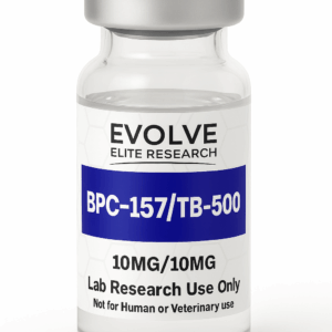
Thymosin Alpha-1 is commonly associated with immune communication and antigen-response research. BPC-157 is more frequently evaluated in tissue-signaling, cellular migration, vascular, and repair-associated models.
Thymosin Alpha-1 is a defined 28-amino-acid immune-research peptide. TB-500 research is generally associated with thymosin beta-4 fragments, actin-related biology, cellular migration, and tissue-response models.
Thymosin Alpha-1 is studied primarily within immune-associated pathways. GHK-Cu is a copper-binding tripeptide investigated in extracellular-matrix, cellular signaling, oxidative, and tissue-remodeling research.
These materials should not be treated as interchangeable. Researchers should select compounds according to the biological question and include appropriate controls when comparing or combining peptides.
Related Research Compounds
Research Resources
Frequently Asked Questions
What is Thymosin Alpha-1?
Thymosin Alpha-1 is a synthetic 28-amino-acid research peptide corresponding to a sequence associated with the N-terminal region of prothymosin alpha. It is commonly studied in experimental models involving immune-cell signaling, dendritic cells, T lymphocytes, cytokines, and pattern-recognition pathways.
What does TA1 mean?
TA1 is a common abbreviation for Thymosin Alpha-1. Scientific literature may also use Tα1 or Thymosin α1.
How many amino acids are in Thymosin Alpha-1?
Thymosin Alpha-1 contains 28 amino-acid residues.
What is the molecular weight of Thymosin Alpha-1?
The reported molecular weight is approximately 3108.30 g/mol.
What is the molecular formula?
The listed molecular formula is C129H215N33O55.
What is the CAS number for Thymosin Alpha-1?
The CAS number is 62304-98-7.
What is the PubChem CID?
The PubChem Compound Identifier associated with Thymosin Alpha-1 is 16132342.
What is the amino-acid sequence?
The sequence is Ser-Asp-Ala-Ala-Val-Asp-Thr-Ser-Ser-Glu-Ile-Thr-Thr-Lys-Asp-Leu-Lys-Glu-Lys-Lys-Glu-Val-Val-Glu-Glu-Ala-Glu-Asn.
Is Thymosin Alpha-1 the same as thymosin beta-4?
No. Thymosin Alpha-1 and thymosin beta-4 are distinct peptides with different sequences, precursor relationships, molecular properties, and areas of research.
Is Thymosin Alpha-1 the same as TB-500?
No. TB-500 research generally involves a thymosin beta-4-related peptide fragment. Thymosin Alpha-1 is a separate 28-amino-acid peptide associated with prothymosin alpha.
What research areas involve Thymosin Alpha-1?
Research areas may include innate immunity, adaptive immunity, dendritic-cell biology, T-cell signaling, natural killer cells, cytokines, Toll-like receptor-associated pathways, antigen presentation, pathogen responses, inflammatory signaling, and cancer immunology.
Does Thymosin Alpha-1 have one confirmed receptor?
Thymosin Alpha-1 is generally studied within complex immune-signaling networks rather than being universally described as a conventional single-receptor ligand. Research has examined Toll-like receptor-associated and other cellular signaling pathways.
Why is Thymosin Alpha-1 studied with dendritic cells?
Dendritic cells coordinate innate sensing, antigen processing, antigen presentation, and communication with T lymphocytes. These functions make them relevant to Thymosin Alpha-1 immune-signaling research.
Why is Thymosin Alpha-1 studied with T cells?
T cells are central to adaptive immunity. Researchers may evaluate whether peptide exposure is associated with changes in T-cell activation, differentiation, proliferation, cytokine production, or regulatory signaling.
Can Thymosin Alpha-1 be used in cytokine studies?
It may be evaluated in controlled laboratory cytokine studies. Researchers should use appropriate untreated, vehicle, stimulated, viability, and time-matched controls.
Can it be studied in pathogen-response models?
Thymosin Alpha-1 has been investigated in experimental models involving host-cell responses to pathogen-associated stimuli. Findings remain specific to the model and measured endpoints.
Can it be studied in cancer-immunology models?
It may be included in controlled experimental research involving immune-cell signaling, antigen presentation, cytokine communication, natural killer cells, T cells, or combination systems.
What does lyophilized mean?
Lyophilized material has undergone freeze-drying to remove a substantial amount of water. This produces a dry material that can offer improved storage stability compared with an experimental solution.
Why can solution stability differ from dry stability?
In solution, peptide molecules have greater mobility and may be more exposed to hydrolysis, oxidation, aggregation, adsorption, enzymes, and microbial contamination.
Why should temperature cycling be minimized?
Repeated temperature changes can introduce condensation, increase environmental exposure, and contribute to inconsistent storage conditions.
What does HPLC evaluate?
HPLC separates detectable components under a defined chromatographic method and can estimate relative sample purity from integrated peak areas.
Does an HPLC result confirm identity?
Not by itself. HPLC evaluates chromatographic behavior and heterogeneity. Molecular identity is better supported with mass spectrometry, sequence analysis, reference comparison, or another orthogonal method.
What does mass spectrometry evaluate?
Mass spectrometry evaluates ion mass-to-charge values and can support identity when the deconvoluted molecular mass is consistent with the expected peptide.
Is HPLC purity the same as peptide content?
No. HPLC purity usually represents the relative area of detected chromatographic peaks. Net peptide content also accounts for water, salts, counterions, and other non-peptide components.
Why are endotoxin controls important?
Immune cells can respond strongly to very small amounts of endotoxin. Without appropriate controls, contaminant-associated activity may be incorrectly attributed to the research peptide.
Why should researchers test multiple concentrations?
A concentration series helps determine whether an observed response is concentration dependent, limited to a specific range, or associated with nonspecific effects at higher concentrations.
Why are time-course experiments useful?
Receptor signaling, gene expression, cytokine production, proliferation, and phenotypic changes can occur on different timelines. Multiple time points provide a more complete response profile.
Why should cell viability be measured?
Viability testing helps distinguish pathway-specific changes from apparent responses caused by reduced cell number, cellular stress, or cytotoxicity.
What is a biological replicate?
A biological replicate is an independently prepared biological sample, culture, biological source, or model system. It measures biological variability rather than repeated measurement of the same sample.
What is a technical replicate?
A technical replicate is a repeated measurement or assay performed on the same biological sample. It helps evaluate procedural and analytical precision.
Why would a researcher select a 10MG vial?
A 10MG vial may be appropriate for multi-concentration experiments, replicate studies, combination research, larger culture volumes, time-course analysis, or workflows requiring material for analytical verification.
Can results from one model be generalized to another?
Not automatically. Results can differ according to species, cell type, concentration, activation state, assay design, exposure duration, and biological environment.
Is this product intended for human use?
No. Thymosin Alpha-1 10MG is supplied strictly for laboratory research use only. It is not intended for human consumption, medical use, veterinary use, household use, or diagnostic use.
Research-Use Notice
Thymosin Alpha-1 10MG is supplied exclusively as laboratory research material. It is not supplied as a drug, finished pharmaceutical product, food, dietary supplement, cosmetic, or consumer product. It is not intended for human consumption, self-administration, medical use, veterinary use, household use, diagnostic use, or therapeutic use.
Scientific information on this page is presented solely in the context of molecular identity, immune-signaling research, cellular and biochemical models, experimental design, and analytical characterization.
No information on this page should be interpreted as instructions for preparation, administration, dosing, self-experimentation, diagnosis, prevention, or treatment of any condition.
This material should be handled only by qualified research personnel in an appropriately controlled laboratory environment. Researchers are responsible for determining suitability for their experimental design and complying with all applicable institutional, local, state, and federal requirements.
Related products
Research Compounds
Research Compounds
Research Compounds
Research Compounds
Research Compounds
Research Compounds
Research Compounds
Research Compounds